
Age-related macular degeneration is the leading cause of certifiable sight loss in the UK. By 2050, the number of people living with AMD is projected to climb past 2.5 million. And in nearly every case, the person who notices something is off first isn’t a hospital consultant — it’s the optometrist who saw them last year for a routine eye test.
That puts independent opticians in a strange position. You’re not the people who treat AMD. But you are, very often, the people who decide whether it gets caught early enough for treatment to matter. With wet AMD, where vision can drop within weeks, that decision sits with you and your referral pathway.
This is a practical guide to building AMD detection, monitoring and referral into how your practice already runs — without bolting on a clinic you don’t have time for, and without losing patients to delayed referrals.
Why AMD belongs in independent practice in 2026
There’s a quiet shift happening in community eye care. Hospital eye services in most parts of the UK are running at or beyond capacity. Wet AMD anti-VEGF clinics in particular are full, and HES departments are leaning more heavily on community optometry to do three things: refer fewer false positives, refer faster on genuine wet AMD, and hold the monitoring load for intermediate dry AMD.
That isn’t a burden — it’s an opportunity. The independent practices doing AMD well in 2026 share a pattern. They’ve turned macular health into a recurring conversation. They have a written pathway. Their OCT capture is consistent. Their referrals get accepted first time. And their patients with intermediate AMD come back every six to twelve months, not because they’re chasing them, but because the practice has made monitoring feel like normal care.
The commercial reality matters too. A practice known locally for catching wet AMD early — and for handling AMD patients with calm, clarity and follow-through — builds the kind of word-of-mouth that no Google Ads spend can buy.
What good AMD detection looks like
“Good” doesn’t mean fancy. It means consistent.
A patient over 55 in your chair should reliably get the same baseline checks every visit: a dilated or undilated fundus image, an OCT scan through the macula if you have one, a careful look at the macula on slit lamp or fundoscopy, and a documented Amsler grid response. Anyone with risk factors — family history, smoker, white European, existing intermediate AMD on the other eye — should get that with no exceptions, even if they’ve come in for a frame repair and you’ve squeezed them into a check.
What pulls practices off course isn’t a lack of kit. It’s drift. The autorefractor used to log a fundus image, but the new technician doesn’t know how. The Amsler grid sits in a drawer instead of being part of the pre-test sheet. OCT is “available” but only ordered when the optometrist asks for it, which means borderline patients slip through.
Good detection is what happens when the workflow runs the same way on a busy Friday with a locum optom as it does on a quiet Tuesday with the principal.
The clinical kit you actually need
You can build a credible AMD pathway with three pieces of equipment.
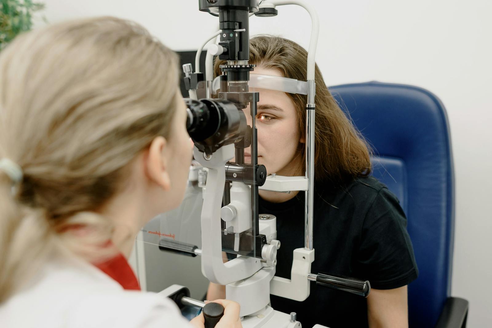
A fundus camera is non-negotiable. Without it you have no longitudinal record of the macula, which means you can’t compare drusen progression year on year. Even a basic non-mydriatic camera is enough for most patients under 70.
An OCT is, by 2026, the difference between a practice that does AMD well and one that doesn’t. It picks up early wet changes — pigment epithelial detachments, intraretinal fluid, sub-retinal fluid — before the patient notices distortion. If you don’t have one, the case to invest is no longer mainly clinical. It’s about referral quality and patient retention.
Amsler grids in printed form, plus a way to record the response in the patient record. Not glamorous. Still essential. Self-monitoring between visits is one of the few things that genuinely catches wet conversion early in the real world.
Anything beyond that — fundus autofluorescence, microperimetry — is a nice-to-have, not a starting point.
The four-step AMD pathway
The independents who run AMD well have a four-step pathway that any team member can follow.
1. Risk-stratify at booking
Anyone over 55, anyone with a family history of AMD, current smokers, and patients flagged from previous visits with drusen or pigment changes — these patients should arrive with a flag on their record. The flag triggers a longer appointment slot, a definite OCT and fundus image, and a printed Amsler grid in the bag they leave with.
The trick here is that the flag has to be set automatically. If it depends on the optometrist remembering, it won’t happen reliably.
2. Standardise the in-chair sequence
Every at-risk patient gets the same sequence: history including any reported distortion, central scotoma, or change in reading vision; Amsler check on each eye; fundus image; OCT macula scan; slit-lamp or volk view of the macula. Same order. Same documentation. Every time.
The reason this matters is comparison. The value of a baseline OCT isn’t the scan itself — it’s the next one, twelve months later, that you can lay alongside it.
3. Decide which bucket the patient is in
After the visit, every AMD-relevant patient should sit clearly in one of four categories:
No AMD, normal age-related changes. Standard recall, mention healthy lifestyle, move on. Don’t over-medicalise.
Early AMD — small drusen, no pigment change, no symptoms. 12-month recall, written information about smoking and diet, baseline imaging captured.
Intermediate AMD — large drusen and/or pigment changes, generally good vision. 6 to 12-month recall, Amsler home monitoring, conversation about AREDS2 supplements, and clear “what to do if vision changes suddenly” instructions.
Suspected wet AMD or significant change. Urgent referral, same day if possible, with imaging attached.
The buckets aren’t original. What matters is that every patient ends up in one, recorded in your system, with a recall and a follow-up plan that matches the bucket. Patients who fall between buckets are the ones who get lost.
4. Refer or monitor — and close the loop
For wet AMD suspicion, the referral has to leave the practice the same day. Most regions now want referrals via electronic eye care referral systems (ERS, NHS e-Referral, or local equivalents). Some HES departments still accept emailed referrals on their template.
Whichever route you use, what the receiving consultant wants is the same: clear documentation of when symptoms started, current vision in both eyes, OCT images, fundus images, and your clinical impression. Vague referrals — “query AMD, please assess” — get rejected or downgraded. Specific referrals — “60-year-old, three-day distortion in right eye, VA dropped from 6/9 to 6/18, OCT shows subretinal fluid, suspect wet AMD, urgent assessment please” — get seen.
For monitoring patients, the loop closes inside your own practice. The recall has to fire. The next visit has to repeat the same imaging. The optometrist has to be able to compare to last year’s scan in under thirty seconds.
How to talk to patients about AMD
This is where independent practices often add the most value — and where the language matters more than the clinical workup.
Patients hear “macular degeneration” and think of going blind. Most won’t say so out loud. They’ll nod, leave, and then quietly stop coming back because the appointment was distressing.
The conversations that work share three features. They’re specific rather than general — “you have some small drusen on the macula, which is very common at your age” beats “you have early signs of AMD.” They separate dry from wet early — “the kind you have is the slow kind, and we monitor it. There’s a different kind we’d act on quickly, and that’s why I’m giving you this grid.” And they end with a clear next action — what to do if you notice change, when you’re back in, and what you can do now (stop smoking, eat well, supplements if intermediate).
The Amsler grid handover is where a lot of practices fall down. Sticking it in the bag isn’t enough. Show the patient how to use it once. Tell them which eye to test first, how often, and what to do if a line bends, breaks or disappears. The single sentence — “if anything looks suddenly different on this grid, ring us the same day” — saves sight in your patient base every year.
Monitoring intermediate AMD between visits
Intermediate AMD is the bucket most independents under-serve. These patients aren’t being referred, and they shouldn’t be — but they need more than a normal recall.
The independents getting this right run something close to a quiet surveillance programme. Six to twelve-month recalls instead of two-year. A standardised “monitoring visit” that’s shorter than a full eye test but always includes OCT macula and fundus imaging. A clinician sign-off comparing to last visit’s images. And a clear note in the record on whether the patient is stable, progressing, or showing any sign of wet conversion.
The cost-of-living squeeze means some of these patients will resist paying for the monitoring visit. The framing that works isn’t “you have AMD, you need this” — it’s “this is how we make sure we catch any change early, because the kind of change we’d act on can happen between full eye tests.” Most patients agree once they understand the why.
For practices that can build it, a small annual subscription that bundles the monitoring visit with the next full eye test removes the friction altogether.
What HES departments actually want in 2026
If your urgent AMD referrals get rejected, downgraded, or sent back for “more information”, the issue is usually the same five things:
Onset isn’t dated. When did the distortion start? Yesterday, last week, last month — this changes the urgency completely. Hospitals are triaging by onset.
Vision isn’t documented in both eyes. Same-day VA, not the one from last year’s test.
OCT isn’t attached. Or it is, but it’s the wrong eye, or it’s a single line scan instead of a macular cube, or the image quality is too low to read.
Symptoms aren’t specific. “Blurred vision” tells the triager nothing. “Sudden central distortion when reading, started Tuesday morning, worse than the other eye” tells them everything.
Your clinical impression isn’t included. Hospitals don’t want you to diagnose for them, but they do want to know what you’re worried about and why. “Suspect wet AMD, possible PED on OCT, urgent assessment” is a useful sentence.
If your practice fixes those five things, your urgent acceptance rate climbs noticeably within a quarter.
AREDS2 and the supplement conversation
For intermediate AMD, the evidence on AREDS2 supplements is strong enough that the conversation should happen at every monitoring visit. The shorthand version: in patients with intermediate AMD in one or both eyes, AREDS2 supplements reduce the risk of progression to advanced AMD by roughly 25 per cent over five years.
That’s not a tiny effect, and patients are entitled to know about it. The conversation that works is brief and honest: “There’s a supplement with strong evidence for slowing this down. It’s not a treatment, and it won’t reverse anything, but the data is good. If you smoke, you need the smoker-safe version. Here’s the brand name, or you can ask a pharmacist.”
Recommending a specific brand sits in a grey area — most practices stick to naming the formulation rather than the product, which keeps you clinically clean.
Where Raven Vision fits
The hard part of running an AMD pathway in 2026 isn’t the clinical decisions. It’s the operational backbone — the flags, the recalls, the image timeline, the referral templates, the audit trail. This is exactly the kind of work a good practice management system should do for you, not pile onto a busy optom or front-of-house team.
The way Raven Vision is set up — built inside Shaukat’s own practices first, opened to other independents after — it handles the moving parts of AMD care specifically: automatic risk flags on the booking diary so at-risk patients get the right slot, structured macular history fields in the clinical record, an image timeline that lets a clinician compare this year’s OCT to last year’s in seconds, templated urgent and routine referral letters with image attachment, and differentiated recall cadences for early versus intermediate AMD.
None of that replaces clinical judgement. It just makes the right thing the easy thing to do.
If you’d like to see how a PMS designed by working optometrists handles this, you can book a demo and we’ll walk through the AMD pathway on a real practice setup, not a sandbox.
Five audit questions every quarter
For practices already running an AMD pathway, the quarterly audit doesn’t need to be elaborate. Five questions tell you whether it’s working:
What percentage of patients over 55 had an OCT macula scan this quarter? (Target: above 85 per cent for at-risk patients.)
How many urgent AMD referrals did we send? How many were accepted on first read? (Target: 90 per cent acceptance.)
How many intermediate AMD patients are on a 6 to 12-month recall, and how many of their recalls were completed on time? (Target: 80 per cent on-time.)
How many monitoring visits did we deliver this quarter, and what’s the trend over the last four quarters? (Direction matters more than absolute number.)
Of the wet AMD referrals we sent, what was the median time from symptom onset to referral? (Target: under five days.)
If you can answer those five questions from your PMS in under ten minutes, you have a pathway that’s running. If you can’t, the pathway exists on paper but not in practice.
Three things to start this month
If you’re reading this and feeling that your AMD detection is more ad-hoc than systematic, you’re not unusual. Almost every independent practice we speak to is somewhere on the spectrum from “we handle it as it comes up” to “we have a full pathway”.
The fastest way to move along that spectrum:
Pick a single age cut-off (55 or 60) and make OCT macula plus fundus imaging mandatory at every visit for everyone above it. No exceptions.
Print Amsler grids, put them at the front desk, and make handing one to every intermediate AMD patient a fixed step in the dispense workflow — not the optometrist’s job.
Audit your last ten urgent AMD referrals against the five things HES wants. Wherever they fell short, fix the template so the next ten don’t.
None of those need new equipment, new staff, or new software. They need consistency. And consistency, in 2026, is what separates the practices that catch wet AMD in time from the ones that hear about it after the fact.
If you’d like to see how this all sits inside a practice management system built for UK independents, book a demo with Raven Vision. We’ll show you what an AMD pathway looks like when the operational side runs itself.