
Most of your patients have never had their blood pressure checked this year. Not because they’re avoiding it — because nobody’s asked. Their GP surgery is booked out for anything that isn’t urgent, the last well-man check was two jobs ago, and the pharmacy machine in the corner of Boots doesn’t count in anyone’s mind as “getting checked.” Then they sit down in your chair for a routine eye test, and you look straight at the one part of their body where high blood pressure leaves a paper trail nobody can talk their way around.
Hypertensive retinopathy doesn’t get anything like the attention diabetic retinopathy does in day-to-day practice conversation, but the arithmetic is similar. The British Heart Foundation and NHS both put the number of adults in the UK living with high blood pressure somewhere north of a quarter of the population, and a large chunk of them don’t know it. Some of those patients are sitting in your chair right now, and the fundus is often the first place the disease becomes visible.
The Eye Test Nobody Realises Is a Blood Pressure Check
Ask a patient when they last had their blood pressure taken and you’ll get a shrug more often than a number. Hypertension is quiet. It doesn’t announce itself with symptoms until it’s already done damage — to the kidneys, the heart, the brain, or the retina. That’s exactly why an eye test matters here: you’re looking directly at small vessels that behave like small vessels everywhere else in the body, without needing to cut anyone open to see them.
This isn’t a new idea in ophthalmic practice — hypertensive changes at the fundus have been part of the clinical vocabulary for over a century. What’s changed is the opportunity. Independent practices are seeing patients more regularly than most GPs manage to, especially since eye tests are one of the few “free” health checks the average working adult still books without being chased. If you’re not treating a chance sighting of arteriolar narrowing or AV nicking as a genuine clinical finding worth acting on, you’re leaving a public health function on the table that very few other places in a person’s life are covering.
What Hypertensive Retinopathy Actually Looks Like
The signs build in a fairly predictable sequence, though not every patient marches through every stage, and plenty sit at a plateau for years on treated hypertension.
The early, easy-to-miss changes
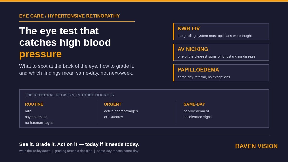
Generalised or focal arteriolar narrowing is usually the first thing you’ll pick up, and it’s the one most likely to get waved through as “just how this patient’s vessels look.” Increased arteriolar light reflex — the vessels start to look shinier, sometimes described as copper wiring and, further along, silver wiring — follows as the vessel walls thicken. Arteriovenous nicking, where a thickened arteriole compresses the vein it crosses, is one of the more reliable signs of longer-standing disease and is worth a deliberate look at every crossing point in the posterior pole, not just the ones your eye lands on first.
The signs that mean something’s actively happening
Flame-shaped haemorrhages, cotton wool spots, and hard exudates — sometimes arranged in a macular star pattern — signal a more active, more recent process rather than years of chronic remodelling. These are the findings that should make you slow down and check the blood pressure history properly, because they suggest control has slipped, or the patient has never actually been diagnosed.
When it’s an emergency, not a referral letter
Optic disc swelling in the context of these vascular signs is the one finding that changes everything about your next move. Papilloedema alongside flame haemorrhages and cotton wool spots points to accelerated or malignant hypertension — a genuine emergency, with real risk of stroke, retinal vein occlusion, and organ damage elsewhere in the body within hours to days. This is not a “book them in with their GP next week” finding. It needs same-day medical assessment, and in most parts of the UK that means straight to A&E or an urgent same-day GP slot arranged by phone while the patient is still with you, not a letter posted that evening.
Grading It Without Overcomplicating the Record
The Keith-Wagener-Barker system from 1939 is still the one most optometrists were taught, running from Grade I (mild arteriolar narrowing) through to Grade IV (papilloedema plus haemorrhages and exudates). In day-to-day practice, most UK clinicians now lean on a simpler three-tier version — non-malignant mild, non-malignant moderate, and malignant — because it maps more cleanly onto the decision that actually matters: does this go to the GP routinely, or does it go somewhere today.
Whichever system you use, the point of grading isn’t to produce a tidy label for the record. It’s to force a decision. A note that says “mild arteriolar attenuation, no haemorrhages, no disc swelling — routine referral to GP for BP check” tells the next person exactly what you saw and what you did about it. That’s worth more than a perfectly accurate KWB grade sitting in isolation with no follow-up action attached to it.
The Referral Decision: Routine, Urgent, or Same-Day
Three buckets cover almost everything you’ll see.
First-detected, asymptomatic, mild changes
Arteriolar narrowing and AV nicking with no haemorrhages, no exudates, and a patient who feels perfectly well — this is a routine referral to their GP for a blood pressure check, written clearly enough that a receptionist triaging incoming letters understands it isn’t urgent. If the patient is already on treatment for hypertension, it’s still worth flagging; it may mean control isn’t as good as their last GP reading suggested, and that’s useful information for whoever’s managing their medication.
Moderate signs with haemorrhages or exudates
This tier deserves a phone call to the surgery rather than a letter sitting in an admin queue for a fortnight. You don’t need to diagnose accelerated hypertension to justify urgency — you need to communicate that these findings are recent and active, not longstanding and stable.
Papilloedema, or anything that reads as accelerated hypertension
Same-day. Phone the practice, phone 111, or send the patient to A&E depending on how unwell they look and how quickly you can get someone else on the line. Document the time you made contact and who you spoke to. This is the one scenario in this whole list where being wrong in the direction of over-caution costs you nothing, and being wrong the other way can cost a life.
Where Hypertension and Diabetes Overlap at the Back of the Eye
A lot of your hypertensive retinopathy patients will also have diabetes, and the two conditions produce overlapping signs — cotton wool spots and haemorrhages appear in both, and distinguishing which disease is driving what you’re looking at isn’t always possible from the fundus alone. We covered the diabetic side of this in more depth in our piece on diabetic eye care — worth reading alongside this one if you haven’t already, because the two pathways in your practice should probably share a lot of their referral logic rather than running as two separate systems in your head.
The practical takeaway: when you see mixed signs in a patient with known diabetes, don’t assume it’s “just” diabetic retinopathy and skip the blood pressure conversation. Hypertension accelerates diabetic retinal damage, and a patient whose diabetic retinopathy seems to be progressing faster than expected is exactly the patient worth asking about their last BP reading.
Making This Routine Without Slowing Down Your List
None of this works if it depends on you remembering to think about it on a busy Tuesday. The practices that catch hypertensive retinopathy consistently, rather than occasionally, have built two small habits into how they run a test.
First, a structured field in the record for vascular findings — not buried in a free-text note that nobody reads back six months later, but a specific, searchable entry. In Raven Vision, that’s a dedicated clinical findings field you can pull a report against, so if you want to check how many BP-related referrals you’ve made this quarter, it’s a filter, not an afternoon of reading old notes. The clinical record system is built so fundus findings, referral actions, and outcomes sit together against the patient, rather than scattered across a paper referral pad and a diary note.
Second, a recall trigger for anyone flagged with early, non-urgent changes who you’re not referring today but want to keep an eye on. If a patient declines a GP referral — and some will, because “my optician thinks I might have high blood pressure” doesn’t always land as urgent to someone who feels fine — that’s a case for a structured six-month recall rather than a hope that they’ll mention it next time they happen to book in. Raven Vision’s recall system handles that kind of clinical follow-up the same way it handles routine two-year recalls, so nothing depends on someone’s memory.
Talking to the Patient
Telling someone you’ve spotted signs of high blood pressure in their eyes is a strange thing to hear from an optician, and how you say it matters almost as much as what you’ve found. Lead with what you actually saw, not a diagnosis you’re not making. “I’ve noticed some changes in the small blood vessels at the back of your eye that can be linked to blood pressure — I’d like you to get it checked with your GP” is honest, specific, and doesn’t put you in the position of diagnosing hypertension from a fundus view alone.
Patients sometimes push back — “my blood pressure’s always been fine” is a common reflex answer. It’s worth a gentle “when was it last actually taken?” in response, because plenty of people are working from a reading that’s three or four years old. Write down what you told them, in their own record, in plain terms. If there’s ever a question later about whether the conversation happened, that note is what backs you up.
A Simple Practice Policy Worth Writing Down
You don’t need a lengthy SOP for this. Three lines, agreed with everyone who tests, and put somewhere the whole team can see it: what counts as routine (mild, asymptomatic, no haemorrhages — GP letter), what counts as urgent (moderate signs, active haemorrhages or exudates — phone call same week), and what counts as an emergency (papilloedema, or anything that looks like accelerated hypertension — same-day, full stop, no exceptions for a busy list). Agree it once, put it on the wall by the slit lamp, and you’ve removed the moment of hesitation that costs the most time when it matters least — right when you’re deciding what to do about a genuinely urgent finding.
If your practice management software makes it easy to tag these findings, pull a recall list, and keep the referral trail against the patient record rather than in three different places, that policy runs itself. If it doesn’t, you end up relying on individual memory for something that should be a system. Raven Vision was built inside working UK practices for exactly this kind of clinical workflow — take a look at pricing or the current offers page, or get in touch to see how the clinical record and recall tools would sit against your own referral pathway.