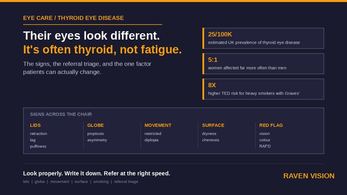

A patient comes in for a routine check and mentions, almost in passing, that people keep telling them they “look tired” or “look surprised” lately. Their prescription hasn’t shifted much. Their vision’s fine. But when you actually look — really look, past the autorefractor number — one eyelid sits a fraction higher than it should, there’s a rim of white showing above the iris that wasn’t there at their last visit, and the eyes don’t quite move together on the way to the corner of the room. Nothing about that shows up on a Snellen chart.
That combination is thyroid eye disease until proven otherwise, and it’s one of the few conditions where an optician is genuinely likely to be the first person to notice — often before the patient’s own GP, and sometimes before the patient even knows they have a thyroid problem at all.
What Thyroid Eye Disease Actually Is
Thyroid eye disease (TED), also called Graves’ orbitopathy, is an autoimmune condition where the immune system turns on the fat and muscle tissue behind the eye rather than the thyroid gland itself. That causes swelling and, over time, scarring of the muscles that move the eye and the fat that cushions it, which is why the classic picture involves the eyes appearing to bulge forward, the lids sitting in the wrong place, and eye movement becoming stiff or restricted. The College of Optometrists puts UK prevalence at roughly 25 per 100,000 people — not common in the way dry eye or blepharitis is common, but not rare enough that an independent practice testing a few thousand patients a year should expect to go without seeing it.
The Graves’ link, and why it’s not automatic
TED overwhelmingly shows up alongside Graves’ disease, the autoimmune cause of an overactive thyroid. UK thyroid charity TEDct notes that more than half of people with an underlying thyroid condition go on to develop some degree of eye involvement, and that a large proportion of those cases go undiagnosed as TED specifically — often written off as tiredness, allergies, or “just how their eyes are.” What’s easy to miss in practice is that the eye disease and the thyroid disease don’t always move in lockstep. Ocular signs can appear before Graves’ is diagnosed, after treatment has already brought thyroid hormone levels back to normal, or — less often — in someone whose thyroid blood tests currently look entirely unremarkable. Waiting for a patient to mention a thyroid diagnosis before you take eye changes seriously is exactly the gap that lets early TED go unrecognised.
The Signs You’ll Actually See Across the Chair
Most of what points to TED is visible without any special equipment — it’s a matter of looking properly and comparing what you see against old photographs or previous notes, which is precisely why a record that includes images is worth more here than a paragraph of free text.
Around the lids
Upper lid retraction is the single most recognisable early sign — the lid sits higher than normal, giving the classic “staring” look, sometimes with visible sclera above the iris (a sign patients themselves often describe as “my eyes look wider” or “more open” than usual). Lid lag on downgaze, puffiness or fullness above and below the eyes, and redness along the lid margin often accompany it.
Around the globe
Proptosis — the eye pushed forward in the socket — can be subtle enough that it only becomes obvious when you compare both eyes side by side or check against a photo from a year earlier. Asymmetry between the two eyes is a strong clue, since TED frequently affects one eye more than the other, at least at first. Restricted or stiff eye movement, and double vision on certain gaze positions, point to the extraocular muscles themselves being involved.
On the surface
Because lid retraction and mild proptosis both reduce how well the lids close, TED patients often present first with what looks like straightforward dry eye — grittiness, watering, redness, light sensitivity, and discomfort with night driving. Chemosis (a boggy, gel-like swelling of the conjunctiva) is a more specific sign worth flagging rather than treating as generic irritation.
None of these signs is unique to TED on its own. A patient can have dry eye without any thyroid involvement whatsoever. What should raise your suspicion is the combination — lid position changes alongside surface symptoms, or asymmetry that wasn’t there at the last visit — rather than any single finding in isolation.
Smoking Is the One Variable You Can Actually Influence
If there’s a single piece of advice worth giving every patient with suspected or confirmed TED, it’s about cigarettes. The British Thyroid Foundation cites figures that are hard to argue with: smokers with Graves’ disease are around twice as likely to develop TED as non-smokers, that risk roughly doubles again — to around fourfold — in smokers who go on to have radioactive iodine treatment for their thyroid, and heavy smokers face something like an eightfold increase in risk compared with people who don’t smoke at all. Smoking cessation doesn’t just lower the odds of developing TED in the first place; patients who continue smoking respond less well to TED treatment generally, and that disadvantage largely disappears once they stop.
This is one of the few genuinely actionable things you can say in the chair that isn’t “go and see someone else.” Pointing a patient toward their GP, a pharmacist, or the NHS Stop Smoking Service costs you thirty seconds and stands a real chance of changing how their eye disease actually progresses — regardless of what an ophthalmologist eventually recommends.
Grading What You’re Looking At, Without Overstepping
You’re not expected to formally stage TED — that’s an ophthalmology job, usually done against a framework that separates disease activity (how inflamed things currently are — pain, redness, swelling, and how quickly function is deteriorating) from disease severity (how much lasting damage or functional threat is present). What matters for you is a simpler, practical version of the same split: is this stable and mild, is it visibly active and getting worse between visits, or is there any sign that vision itself — rather than just appearance — is under threat. That third category is the one that changes how fast you need to act, and it’s worth being deliberately blunt with yourself about which bucket a patient falls into before you decide what to write in a referral letter.
Three Referral Buckets
Routine: mild, stable signs — early lid retraction, mild asymmetry, surface dryness — with no restriction of eye movement, no double vision, and no change in vision. This goes to the GP with a clear description of what you saw, requesting thyroid function tests if these haven’t already been done, and a referral into ophthalmology or a combined thyroid-eye pathway where one exists locally. Many areas now run joint thyroid-eye clinics that bring endocrinology and ophthalmology assessment together in a single appointment, which is worth mentioning in your letter if you know your local system has one.
Urgent: visibly active or worsening disease — increasing proptosis between visits, new or worsening double vision, restricted eye movement, or a rapid change in lid position — warrants a phone call to the eye clinic rather than a routine letter that might sit in a pile for weeks. Active TED can progress meaningfully over a matter of months, and the window for treatments that actually reduce inflammation is time-limited.
Same-day / emergency: two specific pictures justify treating TED as an eye emergency. The first is compressive optic neuropathy — the eye disease pressing on the optic nerve itself, which happens in a small minority of TED cases but can cause permanent vision loss if missed. Watch for reduced vision, dulled or “washed out” colour vision, a relative afferent pupillary defect, or a new visual field defect. The second is corneal exposure from lagophthalmos severe enough that the eye can’t close properly, risking exposure keratopathy and, in bad cases, corneal ulceration. Either picture is a same-day phone call to the eye clinic, not a letter — say plainly why you’re calling, and document who you spoke to and when.
Why Your Records Are the Difference Between a Referral That Lands and One That Doesn’t
TED is a slow-moving condition tracked over months, and a referral letter written from memory a week after the appointment is a weaker document than one written against a structured record taken at the time. What helps most: a dated note of exactly what you saw (lid position, any visible asymmetry, eye movement, surface findings), and — where you have the means — a photograph. A side-by-side comparison with a photo from six or twelve months earlier does more to demonstrate genuine progression than any amount of descriptive prose, and it’s the kind of evidence an ophthalmology triage clerk can act on quickly.
This is where a proper clinical record system earns its keep rather than a free-text box that gets skimmed once and forgotten. In Raven Vision, clinical findings sit as structured, searchable entries against the patient rather than buried in a paragraph, and photos attach directly to the record they belong to — so pulling up “what did this eye look like a year ago” takes seconds rather than a search through old paper notes. It won’t grade the disease for you and it isn’t a substitute for exophthalmometry or imaging done by a specialist — that’s not what it’s for. What it does is make sure the observation you made in the chair is still findable, comparable, and referral-ready months later, which is the part that actually tends to go missing.
The Follow-Up Problem Nobody Plans For
A single referral isn’t the end of the story. TED can flare over months even once a patient is under specialist care, and plenty of practices lose track of these patients between the referral letter going out and whatever happens next — because nothing in the system prompts anyone to check back in. A short-interval recall, three to six months out, for anyone you’ve flagged with active or borderline signs gives you a structured reason to compare photos again and check whether anything’s changed, rather than relying on the patient to mention it unprompted at their next routine test in two years’ time. Raven Vision’s recall system handles that kind of clinical follow-up the same way it handles a standard two-year recall — it’s a list you can build once and let run, not a note you have to remember to act on yourself.
Talking to the Patient
How you raise this matters as much as what you’ve spotted. You’re not diagnosing Graves’ disease, and you shouldn’t sound as though you are. Something like, “I’ve noticed a couple of changes around your eyes that can sometimes be linked to how the thyroid gland is working — I’d like to get this properly checked” is honest, specific, and keeps you in your lane. Patients are often relieved rather than alarmed once they realise there’s a name for what they’d already noticed but couldn’t explain — the “why do I look surprised all the time” question answered at last. Write down what you told them, in plain language, in their own record. If the referral takes a while to be seen, that note is what shows the conversation happened and when.
A Short Practice Policy Worth Having
You don’t need a lengthy protocol for something you’ll see rarely. Three lines, agreed by everyone who tests and kept somewhere visible: what counts as routine (mild, stable, no vision change — GP letter requesting thyroid function tests plus ophthalmology referral); what counts as urgent (active or worsening signs, new double vision — phone the eye clinic that week); and what counts as an emergency (reduced or altered colour vision, an afferent pupillary defect, a visual field defect, or an eye that won’t close — phone the eye clinic today, no exceptions). Agreeing it once removes the hesitation that costs the most time exactly when a case turns out to be the urgent kind.
If your practice management software makes it easy to tag a finding, attach a photo, and set a recall against the same patient record, that policy runs itself in the background. If it doesn’t, you’re relying on someone’s memory for something that should be a system. Raven Vision was built inside working UK practices for exactly this kind of clinical workflow — take a look at pricing or the current offers page, or get in touch to see how the clinical record and recall tools would sit against your own referral pathway. It’s the same underlying habit that catches hypertensive retinopathy early — look properly, write it down properly, and give the next clinician in the chain something they can actually act on.